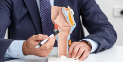
Unlike an adult’s airway which is at its narrowest at the level of the vocal cords, a child’s airway is narrowest in the area below the voice box. At birth the airway in a normal baby is only 5-7mm wide; in premature or low birth weight babies it may be even less than this.
There is not a lot of space in a baby’s airway and any swelling can reduce the space further and cause breathing difficulties.
Stridor
Stridor is the name doctors give to the noisy breathing caused by a partially blocked airway. It is a worrying sign that must always be taken seriously especially in a child. It sometimes sounds like a whistling sound emerging from the chest and is sometimes mistaken for wheezing.
What Causes Stridor?
The cause often depends upon when the child develops the noisy breathing.
From birth
Laryngomalacia
Subglottic stenosis (congenital and acquired)
Congenital voice box abnormalities (cysts, haemangiomas, webs etc)
Recurrent laryngeal nerve palsy
Childhood
Inhaled foreign body
Croup
Epiglottitis
Papillomatosis
Infective causes of stridor
Infections account for a high proportion of cases of paediatric stridor. They tend to happen in early childhood and may occur in epidemics. The two most common causes are croup and epiglottitis.
Laryngomalacia
Laryngomalacia is the most common cause of stridor in small babies. Some babies are born with a voice box that is more “floppy” than usual. When they breathe the cartilages of their voice boxes have a tendency to collapse inwards causing narrowing.
It is usually a mild condition causing noisy breathing which is worse when the baby lies on its back and when it is upset. Most of the time the breathing returns to normal by itself before the baby’s first birthday. In a few children it leads to difficulty with feeding and a failure to thrive.
Diagnosis of laryngomalacia is usually straight forward. A small thin camera or endoscope is passed into the baby’s mouth and the voice box is observed while the baby is breathing, this usually shows the characteristic collapse clearly. Most children grow out of this problem by themselves. Only very few require an operation to improve their breathing. Ary-epiglottoplasty is carried out under general anaesthetic and is a simple procedure that cuts the tight bands closing the voice box.
Sub-glottic stenosis
Narrowing of a child’s airway is most dangerous when it affects the voice box below the vocal cords an area called the sub-glottis. It most often happens because of scarring but sometimes babies are born with a narrow sub-glottis (congenital sub-glottic stenosis). We now know that most cases occur in premature babies who have to have a breathing tube inserted because of the immaturity of their lungs. This life saving treatment can unfortunately lead to scarring in the delicate and narrow area of the sub-glottis.
Congenital narrowing causes breathing difficulties and stridor shortly after the baby is born. As the baby and their airway grow the problems often get better. Acquired stenosis should be suspected in any child who gets stridor and has a previous history of intubation. Often these children do not develop stridor until they are older. They may get recurrent bouts of stridor whenever they get a cold; this is often mistakenly thought to be croup.
In order to diagnose sub-glottic stenosis it is necessary to examine the airway and voice box under an anaesthetic (Microlaryngoscopy and Bronchoscopy).
Many mild cases will settle with further growth. More severe cases may require surgical widening of the airway, called a laryngo-tracheal reconstruction (LTR).
Paediatric Vocal Cord Palsy
The vocal cords are controlled by a pair of important nerves called the Recurrent Laryngeal Nerves. These nerves have a roundabout way of reaching the voice box. They arise in the brain, travel through the neck and then loop back around in the chest to reach the voice box from below. One or both of these nerves may stop working in a young baby. If both nerves are affected then the vocal cords cannot move apart when breathing and stridor will be heard. This usually starts shortly after birth.
The baby may also have an abnormal cry and choking episodes. The paralysis may be temporary and is often blamed on the nerve being pinched in the neck during birth. However, in some children it is associated with more widespread abnormalities of the brain and spine seen in conditions such as the Arnold-Chiari malformation.
Diagnosis & Treatment
An endoscope passed through the mouth shows the narrowed and paralysed vocal cords. An MRI scan is carried out to look for underlying brain and spine problems. Unfortunately, most children in whom both nerves are paralysed will develop severe stridor and breathing difficulties. The only way to treat this is to place a breathing tube below the level of the paralysed vocal cords, this is called a tracheostomy. Once the airway has been made safe the child is observed. Many cases will resolve within 18 months. If there has been no improvement at 2 years the abnormality is likely to be permanent. For the small number of children who do not recover function the aim of surgery is to remove the tracheostomy. Various procedures are used to “lateralise” one vocal fold and widen the airway. Unfortunately, any improvement in the airway is often at the expense of a hoarse and husky voice.
Inhaled Foreign Body
Young children love to put objects into their mouth. Until they are 5 years old children do not have the co-ordination, table manners, or the teeth to chew as well as adults. One of the unfortunate effects of this is that young children are much more likely to choke on or inhale toys, coins, batteries and pieces of food. The choking event may be obvious with choking, retching, and coughing or, unlikely as it may seem, the event may go unnoticed.
There is sometimes a period of stridor and the child may become blue. Paradoxically by the time the child is seen by a doctor they may seem quite well. Rarely there are obvious signs of increased breathing effort and loud and obvious stridor.
Late presentations do occur and the initial difficulty may be misdiagnosed as croup. Obstruction by the foreign body may later produce a chest infection, with low oxygen levels, fever and shivering.
Most often the object lodges in the airways in the chest. Diagnosis
It is important that in any small child with breathing difficulty the possibility of an inhaled foreign body is considered and ruled out. X-rays of the chest are often obtained. These may show the foreign body lodged in the lung or may only show the results of blockage of the lower airway i.e., pneumonia and partial lung collapse.
Treatment
All adults looking after small children should be aware of how to treat a choking attack.
When a child is seen in hospital and there is a suspicion that they may have inhaled a foreign body, the safest approach is often to examine the airway whilst they are asleep. This is called a rigid bronchoscopy.
Croup (laryngo-tracheo-bronchitis) Background
This is a variable illness caused by an infection with a virus. It is most often a mild condition and only around 5% of patients need to be admitted to hospital.
The child is usually aged between 18 months and 2 years. There is often a 2-3 day history of a cold like illness. The most striking thing about croup is the characteristic cough. It is barking and often sounds like a seal. Coughing fits may be followed by episodes of high-pitched stridor. These are usually not severe enough to cause respiratory distress. The child is usually well between coughing/stridor attacks. The illness may last for 1-2 weeks.The key to the diagnosis lies in hearing the characteristic cough. Most children can be looked after at home. The following treatments may be recommended
Humidification - Steam inhalation is unproven but usually advised.
Steroids - More severe cases are treated with steroid medicines.
Intubation - Very rarely breathing difficulties mean that the child needs help with breathing and a tube is placed through the mouth into the lungs to help with this. The child would be given medicine to sedate them and they would be looked after in an intensive care unit.

 close
close